


Ultrasound Technology: How Does It Work
Our highly skilled specialist sonographers carry out all pregnancy and gynaecological scans using the latest in ultrasound technology. We provide 3D and 4D video images, as well as Doppler imaging, which offer the highest levels of accuracy when examining the internal organs and, during pregnancy, the structure of a baby's anatomy and its development.

How does ultrasound technology work?
High frequency sound waves that cannot be heard by the human ear are known as 'ultrasound'. When we carry out scans, we make use of these high frequency waves, which are not harmful to you or your baby.
A small probe - either placed on your skin or gently inserted in your vagina - transmits short pulses of ultrasound into the body. Within a fraction of a second the probe receives multiple “echoes” from the region being examined.
These “echoes” are converted into detailed images of your baby or internal organs and tissue, which your sonographer views on a screen. The use of a gel enhances the transmission of the high frequency waves and the quality of the images that are obtained.
What are the different types of ultrasound scans?
2D and 3D scans: We use 2D and 3D ultrasound scanning technology for pregnancy scans, as well as to scan organs and tissues for diagnostic purposes. 3D gives our sonographer higher resolution visuals than 2D scanning alone, and allows for more accurate diagnosis. Unlike the NHS, all our pregnancy and gynaecology ultrasound scans include 3D/4D technology as standard.
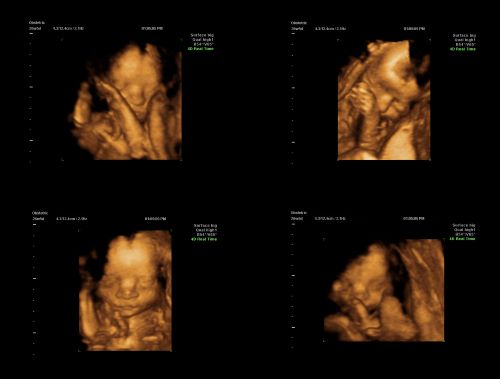
3D pregnancy ultrasound scanning allows us to capture highly detailed images of your baby's internal organs, face, arms and legs, and pick up subtle markers that could indicate chromosomal abnormalities. Our clinic is equipped with the latest technology, including 'HD Live', which can produce stunning static images of unborn babies.
4D pregnancy ultrasound scanning: These are moving video images in 3D, showing your baby's external features more clearly, including the face. We offer both 3D and 4D images at any time during your pregnancy - including the early pregnancy scan, the 12-week nuchal translucency scan, and the 20-week anatomy and anomaly scan.
However, if you’d like to have 3D or 4D images of your baby's face, these will be clearest between 22 and 30 weeks when your baby is bigger and has more fat and muscle, and their facial features are more well-defined.
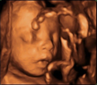
Source: Radiopaedia (https://doi.org/10.53347/rID-60110)
What are Doppler imaging scans?
Our specialists use Doppler imaging including duplex and colour Doppler during all ultrasound scans. This enables us to examine blood flow through the organs and major blood vessels. It is useful in pregnancy to observe placental function and also in gynaecological scans, especially to check ovarian function.
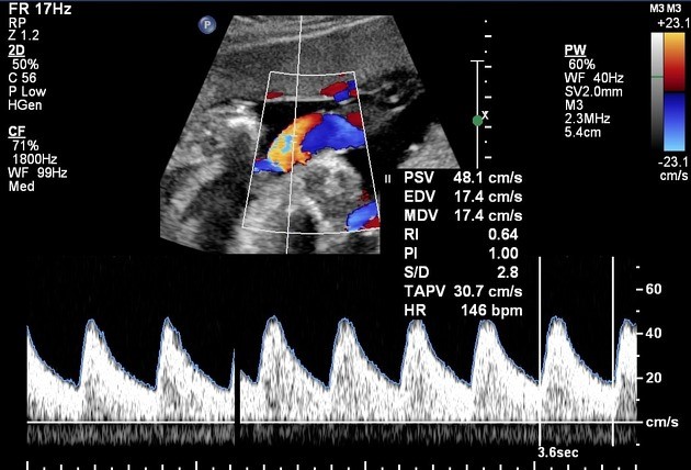
Source: Radiopaedia (https://doi.org/10.53347/rID-37831)
Make an appointment for your private ultrasound scan
If you would like to visit our clinic for a pregnancy or gynaecology scan, please call 020 7244 4200 or make an appointment online.


